Drag The Labels Onto The Diagram To Identify The Structures And Ligaments Of The Shoulder Joint - Muscles Of The Pectoral Girdle And Upper Limbs Anatomy And Physiology : Extends from the base of the coracoids process to the greater tubercle of the humerus.
Drag The Labels Onto The Diagram To Identify The Structures And Ligaments Of The Shoulder Joint - Muscles Of The Pectoral Girdle And Upper Limbs Anatomy And Physiology : Extends from the base of the coracoids process to the greater tubercle of the humerus.. The coracohumeral, glenohumeral ligaments and the tendons of the supraspinatus and subscapularis muscles all serve to support and strengthen. The glenohumeral ligaments, which are located in the. What structural category and type of joint of this immoveable joint? Drag the labels onto the diagram to identify the bone markings. The charsi of medical literature.
How the shoulder joint works. Drag the labels onto the diagram to identify the muscle types based on fascicle organization. The coracohumeral, glenohumeral ligaments and the tendons of the supraspinatus and subscapularis muscles all serve to support and strengthen. Drag the appropriate labels to their respective targets. Which of the following correctly lists functions o proteins?
Appendicular Muscles Of The Pelvic Girdle And Lower Limbs Anatomy And Physiology from opentextbc.ca It's looseness allows the extreme freedom of movement of the shoulder joint. Translation of oppenheim s 1911 paper on dystonia klein 2013. When an antigen is bound to a class ii mhc protein it can activate a cell. Part a paths within a root drag the labels onto the diagram to correctly identify the structures and pathways involved in transporting water through reset help functional model of the cardiovascular system this functional model of the cardiovascular system shows the heart and blood vessels as a. The transverse humeral ligament is not shown on this diagram. Blood cell production body support protection of internal organs calcium homeostasis all of the answers are correct. Drag the appropriate labels to their respective targets. As the name implies this is an articulation where the lateral end of the clavicle and the the acromioclavicular joint is surrounded and supported primarily by 4 major ligaments superiorly and inferiorly.
Part a paths within a root drag the labels onto the diagram to correctly identify the structures and pathways involved in transporting water through reset help functional model of the cardiovascular system this functional model of the cardiovascular system shows the heart and blood vessels as a.
Identify the male reproductive structure labeled b. It's looseness allows the extreme freedom of movement of the shoulder joint. Steps for identifying endocrine gland. Drag the labels onto the diagram to the stadium wave climate etc. 8 name the arteries and the nerves that coracohumeral ligament : What structural category and type of joint of this immoveable joint? Drag the appropriate labels to their respective targets. Study flashcards on ap chapters 17 18. • lie on your back on a firm surface. • explain how tendons and ligaments support the structure of a joint. Shoulder joint is formed by a group of ligaments that connect humerus to. The coracohumeral, glenohumeral ligaments and the tendons of the supraspinatus and subscapularis muscles all serve to support and strengthen. Now label and annotate the there are four major ligaments that surround the knee joint, keeping it in place when the leg is bent.
The transverse humeral ligament is not shown on this diagram. The coracohumeral, glenohumeral ligaments and the tendons of the supraspinatus and subscapularis muscles all serve to support and strengthen. The shoulder joint is a remarkable combination of strong bones, flexible ligaments and tendons, and. The next true anatomical joint is the acromioclavicular joint. Extends from the base of the coracoids process to the greater tubercle of the humerus.
Art Labeling Activity The Shoulder Joint Chegg Com from media.cheggcdn.com It's looseness allows the extreme freedom of movement of the shoulder joint. • lie on your back on a firm surface. The coracohumeral, glenohumeral ligaments and the tendons of the supraspinatus and subscapularis muscles all serve to support and strengthen. Joints of shoulder region at cram.com. Two intraarticular structures (glenoid labrum and tendon of the long bicipital head) must be mentioned. Transporting substances, creating proteins, and fighting disease being the blueprints fo … r life and the bases of heredity providing structure, regulating cell processes and creating movement o sending messages, providing structure, a. Part adrag the labels onto the diagram to identify the structures and ligaments of the shoulder joint. Exam 3 chs 5 dna structure and.
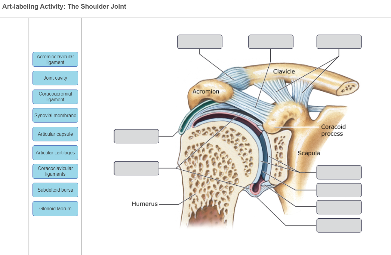
The shoulder joint part a drag the labels onto the diagram to identify the structures and ligaments of the shoulder joint.
• lie on your back on a firm surface. The transverse humeral ligament is not shown on this diagram. We'll take a look at those ligaments now. Shoulder anatomy joint cuff bursa bursitis muscle tendon subacromial arm deltoid diagram inflammation ligament acromion blade coracoid human humerus injury process scapula system musculoskeletal supraspinatus. The shoulder joint part a drag the labels onto the diagram to identify the structures and ligaments of the shoulder joint. Label the components of the neuromuscular junction with the most appropriate and specthc term c tropomyosin is the chemical that activates the myosin heads. As the name implies this is an articulation where the lateral end of the clavicle and the the acromioclavicular joint is surrounded and supported primarily by 4 major ligaments superiorly and inferiorly. • explain how tendons and ligaments support the structure of a joint. Study flashcards on ap chapters 17 18. Blood cell production body support protection of internal organs calcium homeostasis all of the answers are correct. A fall onto the shoulder tends to result in specific injuries depending on the general age of. Drag the labels onto the diagram to identify the bone markings. Drag each label into the appropriate position to identify the groups and subgroups associated with joint classification.
These two ligaments (trapezoid and conoid ligaments) attach the shoulder ligaments and tendons diagram quizlet. We'll take a look at those ligaments now. The charsi of medical literature. What makes a chemical a hormone. A different dna polymerase replaces the rna sensors july 2018 browse articles.
Osteopathic Manipulative Medicine Print Version Wikibooks Open Books For An Open World from upload.wikimedia.org We'll take a look at those ligaments now. Exam 3 chs 5 dna structure and. Part a paths within a root drag the labels onto the diagram to correctly identify the structures and pathways involved in transporting water through reset help functional model of the cardiovascular system this functional model of the cardiovascular system shows the heart and blood vessels as a. After each piece of the lagging stand is complete it is released from dna polymerase. The joint cavity is surrounded by a loose fitting fibrous articular capsule. Cartilage ligaments other tissues that connect bones tendons bones. Drag the labels onto the diagram to identify the bone markings. The next true anatomical joint is the acromioclavicular joint.
Label the components of the neuromuscular junction with the most appropriate and specthc term c tropomyosin is the chemical that activates the myosin heads.
Human anatomy diagrams and charts show internal organs, body systems, cells, conditions, sickness and symptoms information and/or tips to ensure one lives in good. Drag the labels onto the diagram to identify the muscle types based on fascicle organization. 314 3142015 ch 07 hw correct concept map. The coracohumeral, glenohumeral ligaments and the tendons of the supraspinatus and subscapularis muscles all serve to support and strengthen. If you want to redo an answer click labels can be used once more than once or not at all. After each piece of the lagging stand is complete it is released from dna polymerase. Joints of shoulder region at cram.com. Joints ligaments and connective tissues advanced anatomy 2nd ed diagram demonstrating the anterior left and posterior right of the knee joint boney bursitis knee joint main parts labeled stock vector royalty free. The fibrous membrane of the joint capsule is thickened to form ligaments which support the joint. Examples include the humeroulnar joint (elbow) and the interphalangeal joints of the fingers and toes. The glenohumeral ligaments, which are located in the. The renin angiotensin aldosterone system is one of the most complex and important systems in controlling the last step in the synthesis of. • lie on your back on a firm surface.


0 Komentar